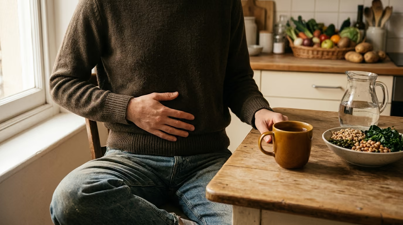

Two muscular sphincters and specialized gastric regions regulate acid secretion, mechanical churning, mucosal protection, and the precise rate of gastric emptying into the intestine.





Many people search online to find out why a meal feels like a heavy weight in their upper abdomen, or how long food actually stays in the stomach. The answers found across wellness forums often portray the stomach as an all-powerful furnace that breaks down and absorbs an entire meal in a single burst. In reality, human physiology operates under a much more coordinated and nuanced set of rules. This guide provides a definitive overview of how the stomach stores, churns, acidifies, and releases food, outlining exactly what this organ does and what it leaves for the rest of the digestive tract.
Medical science views the stomach not as the final site of digestion, but as a biological holding tank, chemical processor, and metered delivery system. The primary work of absorbing nutrients occurs further down in the small intestine. The stomach's true biological purpose is to prepare ingested food so that downstream organs can safely and efficiently handle it. By breaking down large food particles into a uniform slurry and controlling its release, the stomach ensures that the small intestine is never overwhelmed by sheer volume, acidity, or unrefined solids.
Understanding the five core tasks of the stomach helps clarify everyday digestive feelings. A simple way to organize these tasks is to look at how the stomach stores, mixes, acidifies, protects, and meters. When you understand these five steps, common experiences such as post-meal fullness, acid production, and gastric transit become easy to interpret without unnecessary confusion or worry.
The stomach is a hollow, J-shaped muscular organ positioned between the end of the esophagus and the beginning of the small intestine. It is bound by two physiological gates known as sphincters. At the top, the lower esophageal sphincter relaxes to let swallowed food enter while normally preventing acidic fluid from moving upward. At the bottom, the pyloric sphincter serves as a controlled gatekeeper that manages the exit of processed material into the duodenum.
Structurally and functionally, the stomach divides into distinct regions that perform specialized jobs. The upper region, which includes the fundus and the upper portion of the gastric body, acts primarily as a flexible storage reservoir. When you swallow food, vagal nerve reflexes trigger a process called receptive relaxation. This allows the muscular walls of the upper stomach to stretch and expand without a sudden rise in internal pressure, accommodating a meal smoothly.
The lower region of the organ, including the lower body and the muscular antrum, serves as the mechanical engine. Unlike the thin-walled upper reservoir, the antral walls contain thick layers of smooth muscle designed to generate forceful contractions. These muscular contractions sweep downward in regular peristaltic waves, crushing solid food particles against the closed pyloric outlet. This separation of duties ensures that food is stored calmly in the upper section while being processed vigorously in the lower section.
The physical coordination of these regions relies on the enteric nervous system, an intricate network of neurons embedded directly within the stomach wall. This local network communicates constantly with the central nervous system through parasympathetic and sympathetic pathways. Parasympathetic signals, primarily carried by the vagus nerve, stimulate muscular motility and trigger glandular secretions during and after eating. Sympathetic activity, typically elevated during times of physical stress, slows motor contractions and delays gastric processing. Readers interested in the broader intestinal environment can read our overview of digestion and everyday gut function.
Inside the gastric folds, millions of microscopic gastric pits descend into specialized glands that secrete roughly two to three liters of gastric juice each day. Gastric juice is not a single uniform liquid. It is a precise mixture of water, hydrochloric acid, digestive proenzymes, electrolytes, and specialized protective glycoproteins. Each component is manufactured by dedicated cell types that respond to chemical and neural triggers.
Parietal cells, located primarily in the middle regions of the gastric glands, are responsible for secreting concentrated hydrochloric acid. These cells utilize specialized proton pumps to transport hydrogen ions against a massive concentration gradient into the gastric cavity. The resulting acidic environment creates an internal pH typically between 1.5 and 3.5 during active digestion. This high acidity serves two essential roles. It denatures the complex three-dimensional structures of dietary proteins, unfolding them like tangled string, and it destroys a wide variety of ingested environmental microbes.
Chief cells, another key cellular group, synthesize and release an inactive proenzyme called pepsinogen. Pepsinogen remains inactive while inside the chief cell to prevent the cell from digesting its own internal proteins. Once released into the acidic fluid of the stomach lumen, the low pH cleaves a segment of the pepsinogen molecule, converting it into active pepsin. Pepsin is an endopeptidase, an enzyme that cuts internal peptide bonds within protein chains, breaking long proteins into shorter polypeptide fragments.
Gastric secretion is not exclusively focused on protein. Chief cells also secrete gastric lipase, an enzyme that begins the breakdown of dietary triglycerides into diglycerides and free fatty acids. Although gastric lipase handles only a fraction of overall fat breakdown compared to pancreatic enzymes, it plays an important role in initiating fat digestion in an acidic setting. Parietal cells also produce intrinsic factor, a specialized transport glycoprotein that is essential for the later absorption of vitamin B12 in the terminal ileum. To learn more about how dietary components are processed throughout the body, visit our guide on food, fiber and nutrition.
Chemical secretions alone cannot break down a dense meal of solid food. Mechanical reduction is necessary to expose the interior of food particles to gastric acid and enzymes. When solid food enters the stomach, it arrives as chewed boluses mixed with saliva. The stomach converts these irregular solids into chyme, a smooth, semiliquid suspension suitable for the delicate mucosal tissues of the small intestine.
This mechanical breakdown relies on an action called antral retropulsion. Peristaltic contraction rings arise in the upper body of the stomach and travel downward toward the antrum, gaining speed and force as they move. As a peristaltic wave approaches the pylorus, the pyloric sphincter narrows and almost completely closes. When the advancing wave pushes gastric contents against this constricted opening, only a tiny squirt of liquid passes through into the duodenum. The remaining bulk of the solid food is slammed against the closed muscular barrier and propelled forcefully backward into the body of the stomach.
This continuous cycle of forward propulsion, impact, and backward jetting crushes food particles against one another. The sheer physical shear forces generated by retropulsion grind solids down until they are reduced to particles generally smaller than one to two millimeters in diameter. Food particles that remain larger than this threshold cannot clear the pyloric canal and must undergo repeated cycles of grinding.
The physical nature of the meal dictates how long this mechanical processing takes. Liquids pass through the stomach rapidly because they require no mechanical grinding. Fine, soft carbohydrates break down and liquefy much faster than dense, fibrous cuts of meat or high-fat meals. Until solids are reduced to a uniform liquid suspension, the stomach holds them within its churning antral mill. For a wider view of daily gut function and motility patterns, explore our articles on digestive health.
A biological paradox sits at the center of gastric physiology: the stomach produces acid strong enough to corrode metal and enzymes designed to digest meat, yet it does not destroy its own muscular walls. The organ survives this harsh internal environment through a dynamic defense system known as the gastric mucosal barrier. This protective network balances aggressive luminal factors with continuous tissue defense.
The primary physical defense is the mucus-bicarbonate barrier. Specialized surface mucous cells and neck cells continuously secrete an adherent, gel-like mucus composed of cross-linked mucin glycoproteins. This viscous gel adheres tightly to the epithelial surface, forming an unbroken protective blanket that coats every fold and pit. Beneath this mucus layer, surface epithelial cells secrete bicarbonate ions into the gel matrix. The trapped bicarbonate neutralizes hydrogen ions trying to diffuse toward the cellular lining, creating a dramatic pH gradient across a space thinner than a millimeter. While the gastric lumen may have a highly corrosive pH of 2.0, the immediate surface of the living epithelial cells rests in a near-neutral microenvironment around pH 7.0.
In addition to the chemical buffer, the surface epithelial cells are linked together by tight junctions. These tight intercellular seals prevent acid and pepsin from leaking between cells into deeper tissue layers. If minor epithelial damage occurs, the stomach uses a rapid repair process called restitution. Nearby healthy cells detach from the basement membrane and migrate across the damaged area to close the breach within minutes. The entire cellular lining of the stomach is completely replaced every three to five days through rapid cell division in the gastric pits.
Beneath the epithelial layer, a dense microvascular blood supply delivers the oxygen, glucose, and bicarbonate needed to sustain this intense metabolic activity. Local blood flow also carries away any hydrogen ions that manage to penetrate the epithelial surface. This entire defensive cascade is regulated by endogenous prostaglandins, chemical messengers synthesized from dietary lipids that stimulate mucus production, promote bicarbonate secretion, and maintain healthy microvascular blood flow. When factors such as severe physiological stress, chronic inflammation, or nonsteroidal anti-inflammatory drugs disrupt prostaglandin production, this protective balance can weaken.
Gastric emptying is the process by which chyme moves from the stomach into the duodenum. It is not an uncoordinated dump of fluid, but a tightly regulated delivery system. The primary goal of gastric emptying is to deliver nutrients and acid to the small intestine at a steady rate that matches the digestive and absorptive capacity of the downstream organs.
The rate of emptying is controlled by a coordinated feedback loop known as the enterogastric reflex, alongside gut hormones released by the duodenal wall. When acidic, hypertonic, or fat-rich chyme enters the duodenum, specialized receptors detect these signals immediately. In response, endocrine cells in the intestinal mucosa release regulatory hormones, including cholecystokinin, secretin, and gastric inhibitory peptide. These hormones act on the stomach to relax the proximal reservoir, slow antral contractions, and tighten the pyloric sphincter, delaying further emptying until the duodenum neutralizes the acid and processes the incoming fat.
Because of this constant feedback, different meal components leave the stomach at vastly different rates:
In clinical medicine, gastric emptying is evaluated using gastric emptying scintigraphy. In this diagnostic study, a patient consumes a standardized meal labeled with a minute, safe amount of a radioactive tracer, and a gamma camera records its progress over four hours. Cleveland Clinic protocols provide clear clinical test benchmarks for a standard solid meal:
For a specialized liquid emptying test, the normal clearance range is much faster, typically sitting between 20 and 25 minutes. It is vital to recognize that these scintigraphy numbers represent standardized clinical test thresholds under laboratory conditions, rather than a universal guarantee for every meal eaten at home. For readers tracking daily abdominal sensations, our resource on bloating and regularity offers helpful context.
A common misunderstanding about the stomach is the assumption that it extracts and absorbs most of the nutrients in our food. In reality, the stomach's absorptive capacity is limited. While the stomach works tirelessly to store, churn, and denature food, it is anatomically unsuited for large-scale nutrient absorption.
The stomach lacks the massive, folded absorptive surface area found in the small intestine. It does not possess circular folds, villi, or microvilli, which expand the surface area of the intestinal tract by hundreds of times. Furthermore, the thick mucus-bicarbonate barrier and tight intercellular junctions that protect the stomach lining also serve as a barrier to the passage of most vitamins, minerals, amino acids, and carbohydrates. The stomach absorbs only a tiny fraction of ingested components, including water, lipid-soluble compounds, small amounts of alcohol, and certain acidic medications such as aspirin.
The stomach's true biological contribution is preparing food for downstream absorption. By converting solid food into finely ground liquid chyme, the stomach expands the surface area of the meal thousands of times over. When this chyme enters the duodenum, it is met by pancreatic juice containing bicarbonate, which rapidly neutralizes the gastric acid, and an array of powerful digestive enzymes, including trypsin, chymotrypsin, amylase, and pancreatic lipase.
Without the stomach's preparatory grinding and controlled release, the small intestine would struggle to process large, dense food particles, leading to malabsorption, osmotic diarrhea, and severe discomfort. The stomach is an essential upstream partner that makes downstream absorption smooth and efficient. You can read more about overall gut maintenance at DigestGenius.
When the normal mechanical or secretory functions of the stomach are disrupted, specific clinical patterns emerge. One of the most studied functional disorders of gastric motility is gastroparesis, a chronic condition characterized by delayed gastric emptying in the complete absence of a mechanical obstruction.
In clinical practice, establishing a diagnosis of gastroparesis requires three criteria:
The symptoms of gastroparesis can significantly disrupt daily life. According to clinical registry protocols established by the National Institute of Diabetes and Digestive and Kidney Diseases, patients diagnosed with gastroparesis frequently report a distinct cluster of complaints. In registry cohorts, nausea occurs in roughly 90% of patients, vomiting in over 80%, early satiety (feeling full after eating only a few bites) in about 60%, abdominal bloating in around 75%, and upper abdominal pain in approximately 50%.
It is important to remember that these registry statistics reflect individuals with confirmed, clinically managed conditions, rather than the general public. Furthermore, symptoms alone do not prove that a person has delayed gastric emptying. Upper abdominal fullness, belching, and nausea are nonspecific signs that overlap with other common gastrointestinal conditions, including functional dyspepsia, Helicobacter pylori infection, and peptic ulcer disease. Objective diagnostic testing is essential to separate true motility delays from visceral hypersensitivity or mucosal inflammation.
Discussions surrounding stomach acid, digestion time, and gut function are filled with myths. Correcting these misconceptions with physiological facts helps reduce health anxiety and fosters a grounded understanding of the body.
A common belief is that the stomach does all the work of digestion, leaving the intestines simply to clear out the remainder. In reality, the stomach only performs initial mechanical breakdown, begins protein denaturation, and starts minor fat hydrolysis. The bulk of chemical digestion, including carbohydrate breakdown, complete protein cleavage, fat emulsification, and nearly all nutrient absorption, happens in the small intestine.
Many people use the terms "stomach acid" and "digestive enzymes" interchangeably. Hydrochloric acid is an inorganic chemical solution secreted by parietal cells that creates an acidic environment and denatures protein structures. Enzymes, such as pepsin and gastric lipase, are biological protein catalysts produced by chief cells that chemically cut molecular bonds. Acid activates and assists enzymes, but they are entirely different biological tools.
People often search for the exact number of hours it takes to digest food, assuming every meal follows an identical biological timer. Gastric transit is dynamic and depends entirely on the composition of the meal. A glass of water can leave the stomach in less than twenty minutes, whereas a large meal rich in dietary fats, dense proteins, and coarse fiber can take four hours or more to clear the antrum.
Because gastric juice is corrosive, people sometimes assume that any presence of acid causes wear and tear on the stomach wall. Under normal physiological conditions, the stomach maintains a robust mucus-bicarbonate barrier that keeps the tissue surface at a neutral pH. The stomach is built specifically to handle its own acidic secretions safely.
You do not need restrictive regimens or unproven supplements to support your stomach's natural physiology. The stomach operates best when everyday lifestyle choices align with its built-in mechanical and chemical processes.
A grounded, practical lifestyle habit you can adopt immediately is conscious meal pacing and thorough chewing. The stomach's lower muscular mill must reduce solid food particles down to smaller than two millimeters before the pylorus allows them to pass into the duodenum. When you eat in a rush and swallow large, unchewed pieces of food, the antrum has to perform extensive mechanical work, which delays gastric transit and often causes feelings of heaviness.
Taking twenty minutes to finish a meal and chewing each bite thoroughly relieves mechanical strain on the antrum. Furthermore, eating in a calm, seated position allows the vagus nerve to properly regulate receptive relaxation in the upper reservoir, accommodating incoming food without sudden spikes in gastric wall tension.
Scientists are currently investigating the bioelectric rhythms that drive gastric contractions, focusing on specialized pacemaker cells known as the Interstitial Cells of Cajal. These cells generate continuous electrical slow waves that dictate the frequency and direction of gastric peristalsis, typically running at a baseline rate of three cycles per minute in healthy humans. Researchers are studying how disruptions in these electrical signals, termed gastric dysrhythmias, contribute to unexplained nausea and chronic functional dyspepsia. While electrogastrography and bioelectric pacing therapies show promise in clinical trials, their widespread use as standard diagnostic and therapeutic tools is still evolving.
Everyday variations in digestion are normal, but persistent or severe upper gastrointestinal symptoms should not be ignored. You should consult a qualified physician or gastroenterologist if you experience any of the following red-flag symptoms:
These signs require thorough clinical evaluation, which may include upper endoscopy, imaging, or laboratory testing to identify and treat underlying conditions safely.
To support your gastric mechanics and promote comfortable transit this week, try these practical steps:
DigestGenius publishes research-led guidance on digestion, the gut microbiome, fiber, probiotics, gut-brain signaling, inflammation and everyday digestive wellbeing.


Explore clear, research-led guidance on digestion, the microbiome, food, fiber, probiotics and the gut-brain connection.
read the blog