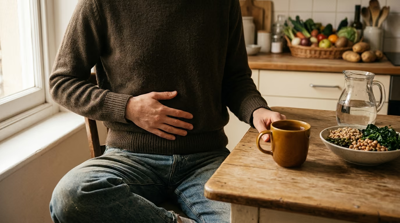

Many people credit the stomach with digestion, but the small intestine actually completes nutrient breakdown and mucosal absorption through an intricately folded surface.





Many people find themselves searching online for explanations when everyday meals cause unpredictable bloating, fatigue, or discomfort. You might wonder why your body feels depleted even when you eat nutrient-dense meals. The answer often lies within the small intestine, the central organ responsible for breaking down food and transferring nutrients into your body.
Understanding how this organ works helps clear away confusing gut wellness trends. This guide provides a definitive, science-based explanation of intestinal architecture, chemical digestion, and nutrient absorption. You will learn how your digestive tract processes everyday foods and what to do when digestion does not go as planned.
Medical researchers and gastroenterologists agree on the primary role of the small intestine. It serves as the primary processing center where chemical breakdown concludes and nutrient uptake occurs. Without this continuous biological process, the human body cannot extract energy, vitamins, or minerals from food.
Scientific consensus divides the work of the small intestine into three synchronized functions. First, it mixes incoming food with digestive fluids from the pancreas, liver, and intestinal lining. Second, it uses specialized cells to absorb broken-down molecules across an expansive surface area. Third, it propels remaining, unabsorbed residues into the large intestine while reclaiming the majority of fluid.
Researchers emphasize that the small intestine does not work in isolation. It relies on coordinated signaling from the stomach, pancreas, liver, and nervous system. Digestion begins in the mouth and stomach, but the small intestine finishes the process and manages systemic delivery.
Health authorities emphasize that nutrient absorption requires both mechanical movement and precise biochemical reactions. When these processes operate smoothly, the body efficiently absorbs carbohydrates, proteins, fats, vitamins, and minerals. If any part of this system is disrupted, nutrient delivery declines and digestive symptoms emerge. To better understand these integrated processes, readers can review broader principles of digestion and everyday gut function.
Physiologists break the overall process of intestinal digestion and nutrient uptake into three distinct phases. These phases occur sequentially as food travels through the digestive tract. They separate the initial breakdown of large food pieces from the final transport of nutrients into the bloodstream.
The first phase occurs directly inside the central cavity, or lumen, of the small intestine. Partially digested food, known as chyme, arrives from the stomach in acidic batches. The small intestine immediately neutralizes this acid using bicarbonate from pancreatic secretions.
Once the environment is neutralized, pancreatic enzymes begin breaking down large food molecules. Pancreatic amylase splits complex starches into smaller carbohydrate chains. Proteases such as trypsin and chymotrypsin cleave long protein chains into shorter peptides. Meanwhile, bile salts from the liver emulsify dietary fats so pancreatic lipase can break triglycerides into smaller fatty acids.
Intraluminal hydrolysis reduces dense food into smaller intermediate molecules. However, most of these intermediate fragments are still too large for the body to absorb. They must undergo further processing at the intestinal surface before they can enter circulation.
The second phase takes place directly on the surface of the intestinal lining. The cells lining the intestine, known as enterocytes, feature microscopic projections called microvilli. Together, these microvilli form what scientists call the brush border.
The membrane of the brush border contains specialized enzymes anchored to the cell surface. These enzymes perform the final chemical cuts on intermediate food molecules. For example, disaccharidases such as maltase, sucrase, and lactase split double sugars into single sugar units like glucose, fructose, and galactose. Peptidases on the brush border split short peptide chains into individual amino acids and small peptide pairs.
Brush-border digestion ensures that nutrients are broken down into their smallest possible components. This localized breakdown occurs right next to transport proteins. As a result, newly freed nutrients can immediately move into the absorptive cells.
The third phase is mucosal absorption, where individual nutrient molecules cross the intestinal barrier. Specialized transporter proteins embedded in the enterocyte membrane pull nutrients inside the cell. Some nutrients move through passive transport, while others require active transport powered by cellular energy.
Once inside the enterocyte, nutrients follow specific pathways depending on their chemical nature. Water-soluble molecules, including single sugars, amino acids, and water-soluble vitamins, move across the base of the cell. They enter nearby capillary blood vessels and travel toward the liver.
In contrast, dietary fats undergo reassembly inside the enterocyte. The cell packages these lipids into specialized transport particles called chylomicrons. Chylomicrons exit the cell and enter small lymphatic vessels called lacteals, bypassing immediate bloodstream delivery.
The small intestine has an intricate physical structure designed to support digestion. It is not a smooth, rigid pipe. Instead, its inner wall contains multiple tiers of folding that maximize the physical contact between intestinal tissue and digested food.
The largest structural feature of the intestinal lining is a series of permanent mucosal folds. These circular ridges are known scientifically as plicae circulares. They project inward toward the passing food stream, particularly within the upper regions of the intestine.
Circular folds serve two essential functions. First, they increase the overall internal surface area of the organ. Second, they act as speed bumps that cause passing chyme to spiral slowly rather than moving in a straight line. This spiraling motion gives digestive enzymes more time to break down food.
Because these folds do not flatten out when the intestine is full, they provide continuous surface amplification. They ensure that passing nutrients remain in constant contact with the intestinal lining.
Projecting from the circular folds are millions of tiny, finger-like structures called villi. Villi extend directly into the fluid stream of the intestinal cavity. Each individual villus acts as a self-contained absorptive unit equipped with its own internal transport network.
Inside every villus lies a web of microscopic blood capillaries alongside a central lymphatic vessel known as a lacteal. The capillary network captures water-soluble nutrients, while the lacteal collects packaged dietary fats. This internal arrangement allows absorbed material to move rapidly away from the lining and into general circulation.
Between the bases of neighboring villi sit small pockets called crypts of Lieberkühn. These crypts house stem cells that continuously divide to produce new intestinal cells. Newly formed cells migrate up the sides of the villus, mature into functional enterocytes, and replace older cells every few days. Readers interested in cellular maintenance can learn more about the gut barrier and immune function.
The smallest layer of surface architecture exists on the individual enterocyte cells. The outer surface of each enterocyte is covered with thousands of microscopic projections called microvilli. Under a microscope, this dense layer looks like the bristles of a brush.
Physiology research indicates that a single enterocyte can feature roughly 3,000 microvilli on its apical surface according to NCBI Bookshelf resources. These microvilli dramatically increase the microscopic membrane area available for nutrient interaction. They also hold digestive enzymes and transport channels in place.
The brush border serves as the final boundary between the outside world and the body's interior. It acts as an active biochemical barrier that selects necessary nutrients while keeping unwanted compounds out.
For decades, popular health articles claimed that the human small intestine had the surface area of a tennis court. Modern scientific measurements have corrected this exaggerated comparison. Morphometric studies published by researchers like Helander and Fandriks provide realistic surface estimates.
Current morphometric evidence indicates that circular folds enlarge the mucosal surface by roughly 1.6 times. Villi and microvilli amplify the surface area by an additional 60 to 120 times according to Helander and Fandriks. When evaluating the total gastrointestinal mucosa from mouth to rectum, the adult surface area measures roughly 30 to 40 square meters.
Other physiological reviews report that intestinal folds, villi, and microvilli collectively expand the surface area between 30-fold and 600-fold depending on measurement methods. Keeping these varying estimates in mind helps prevent misconceptions. The intestinal lining is exceptionally large and efficient, but it does not match the size of an athletic court.
The small intestine measures approximately twenty feet in length in an adult. It is anatomically divided into three distinct segments: the duodenum, the jejunum, and the ileum. Each region possesses specialized structural features tailored to specific digestive and absorptive tasks.
The duodenum is the shortest segment of the small intestine, measuring roughly ten to twelve inches. Despite its short length, it plays a vital role in preparing chyme for absorption. It receives acidic food from the stomach and blends it with bile from the liver and enzymes from the pancreas.
The duodenum neutralizes gastric acid, creating an optimal environment for digestive enzymes. It is also the primary site for the absorption of certain key minerals. Iron absorption occurs predominantly in the duodenum, where enterocytes use specialized transport mechanisms to take up dietary iron.
Because the duodenum handles initial chemical mixing, structural changes or inflammation here can impair overall digestive efficiency. When duodenal function is compromised, downstream segments must work harder to process food components.
The jejunum forms the middle two-fifths of the small intestine. It features tall, densely packed villi and prominent circular folds that maximize absorptive capacity. The vast majority of macronutrient absorption occurs within this highly active segment.
As chyme moves through the jejunum, enterocytes absorb single sugars, amino acids, and small peptides. The proximal two-thirds of the jejunum also serves as the primary site for dietary fat absorption according to gastrointestinal physiology reviews. Blood flow to the jejunum increases substantially after a meal to support rapid nutrient transport.
The jejunum operates with great speed and capacity. By the time food finishes its transit through this middle section, most usable calories, vitamins, and minerals have already entered the body.
The ileum is the final and longest segment of the small intestine, connecting directly to the large intestine. While its villi are shorter and less dense than those in the jejunum, the ileum performs critical specialized tasks. It serves as an essential site for absorbing nutrients that bypassed earlier segments.
The terminal ileum has a specific role in absorbing vitamin B12. This process requires intrinsic factor, a protective protein produced in the stomach that binds to vitamin B12. The terminal ileum also reabsorbs bile salts, recycling them back to the liver through the bloodstream for reuse in future meals.
Additionally, the ileum contains dense clusters of immune tissue known as Peyer's patches. These immune monitoring stations help sample gut contents and protect against harmful bacteria. To learn more about general bowel habits and digestive patterns, explore our guide on bloating and regularity.
Every meal contains a unique mix of carbohydrates, proteins, fats, fluids, and electrolytes. The small intestine uses tailored biochemical pathways to process each of these dietary components. Understanding these pathways clarifies how different foods are digested.
Carbohydrates supply the body with readily accessible energy. Dietary carbohydrates include complex starches, double sugars like sucrose and lactose, and single sugars like glucose and fructose. Chemical digestion of starch begins in the mouth with salivary amylase, pauses in the stomach, and resumes vigorously in the small intestine.
Pancreatic amylase breaks complex starches into smaller maltose units. Next, brush-border enzymes such as maltase, sucrase, and lactase split these units into individual monosaccharides. Enterocytes then absorb these single sugars using specialized transporter proteins.
When brush-border enzymes are insufficient, unabsorbed sugars remain in the intestinal cavity. This can draw water into the bowel and feed colonic bacteria, leading to gas and loose stools. Readers can review our resources on food, fiber and nutrition for deeper dietary context.
Dietary proteins provide the raw building blocks required for muscle repair, enzyme production, and immune defense. Protein digestion begins in the stomach, where gastric acid and the enzyme pepsin denature complex protein structures into loose peptide chains.
Once these chains enter the small intestine, pancreatic proteases continue breaking them down. Trypsin, chymotrypsin, and carboxypeptidase break the chains into short oligopeptides and free amino acids. Peptidases embedded in the brush border perform the final breakdown steps at the cell surface.
Enterocytes absorb both single amino acids and small dipeptides or tripeptides using specialized membrane transporters. Once inside the enterocyte, internal enzymes convert remaining small peptides into individual amino acids. These amino acids then pass into the capillary network and travel via the portal vein directly to the liver.
The digestion and absorption of dietary fats follow a completely different pathway than carbohydrates and proteins. Because lipids do not dissolve in water, they form large fat droplets in the watery fluid of the intestine. The body must break these droplets apart before enzymes can digest them.
Bile salts from the liver attach to large fat droplets, breaking them down into microscopic droplets through emulsification. Pancreatic lipase then hydrolyzes triglycerides into free fatty acids and monoglycerides. These digestion products combine with bile salts to form microscopic clusters called micelles.
Micelles transport fatty acids directly to the brush-border surface, where the lipids diffuse into the enterocyte. Inside the cell, the endoplasmic reticulum reassembles these fatty acids back into triglycerides. The cell packages these triglycerides, along with cholesterol and proteins, into particles called chylomicrons.
Instead of entering the bloodstream directly, chylomicrons exit the enterocyte and enter lacteals. These specialized lymphatic vessels carry the chylomicrons through the lymphatic system. They eventually enter the bloodstream near the heart, allowing dietary fats to circulate before reaching the liver.
In addition to processing solid food, the small intestine manages large volumes of fluid every day. The digestive system processes roughly eight to nine liters of fluid daily from dietary drinks, saliva, gastric juice, bile, and pancreatic secretions.
The small intestine absorbs the vast majority of this fluid along with essential electrolytes such as sodium, potassium, and chloride. Water absorption occurs passively, following the osmotic gradient created by the active transport of sugars, amino acids, and sodium.
According to the Merck Manual Consumer edition, the small intestine absorbs all but about one liter of fluid before passing the remaining contents to the large intestine. This continuous fluid reclamation maintains systemic hydration and prevents severe water loss.
Gastrointestinal biology is often oversimplified in popular health media. These simplifications can lead to confusion regarding diet, digestion, and gut health. Examining common myths alongside biological facts clarifies how the small intestine actually works.
Many people use the terms digestion and absorption interchangeably. In reality, they represent two distinct biological stages. Digestion refers to the mechanical and chemical breakdown of large food pieces into tiny molecules. Absorption refers to the physical movement of those molecules across the cellular lining and into the body.
You can have normal chemical digestion but impaired absorption if your intestinal lining is damaged. Conversely, if your body lacks specific digestive enzymes, absorption cannot occur because food fragments remain too large. Recognizing this difference helps pinpoint the true cause of digestive challenges.
A widespread misconception is that bile acts as a digestive enzyme that dissolves fat. Bile contains bile salts, cholesterol, and phospholipids, but it does not contain digestive enzymes. Its primary role is physical emulsification rather than enzymatic breakdown.
Bile acts like dish soap, breaking large grease globules into microscopic droplets. This physical separation gives pancreatic lipase access to break down fat molecules chemically. Without pancreatic lipase, bile alone cannot complete fat digestion.
It is often claimed that every nutrient you eat enters the portal vein and goes directly to the liver for screening. While this holds true for carbohydrates, amino acids, water-soluble vitamins, and short-chain fatty acids, it is incorrect for dietary fats.
Long-chain dietary fats are packaged into chylomicrons and transported through the lymphatic system. These fats enter general blood circulation before reaching the liver. This distinct pathway explains why dietary fats affect blood lipid levels differently than carbohydrates or proteins.
Many assume that eating enough food prevents nutritional deficiencies. However, nutritional status depends entirely on mucosal absorptive capacity. If the intestinal lining is inflamed or damaged, essential nutrients pass through unabsorbed regardless of dietary intake.
For example, individuals with untreated celiac disease experience immune-mediated damage to their villi according to clinical health sources. This damage flattens the absorptive surface, resulting in malabsorption of iron, calcium, and fat-soluble vitamins despite consuming adequate food.
Older textbooks frequently compared the small intestine to a tennis court, claiming a surface area of over two hundred square meters. While this comparison made the concept memorable, it relied on flawed mathematical models.
Modern morphometric evaluations by Helander and Fandriks confirm that the entire human gastrointestinal mucosa measures between 30 and 40 square meters. The small intestine comprises the majority of this surface, but its true area is far smaller than older estimates suggested.
Examining realistic biological models helps illustrate how the small intestine handles different digestive demands. The following scenarios demonstrate how the organ responds to typical dietary challenges and health conditions.
Consider an illustrative model of a person eating a meal rich in complex starches, such as whole grains and tubers. As the food reaches the duodenum, the pancreas releases amylase to break the starches into oligosaccharides and maltose.
As these fragments move along the jejunum, brush-border enzymes split them into individual glucose molecules. Transporter proteins move the glucose into enterocytes, which release it into villus capillaries. The portal vein transports this glucose to the liver, which regulates blood sugar release. This scenario illustrates how luminal enzymes, brush-border proteins, and vascular networks work together during normal digestion.
In an illustrative scenario involving lactase insufficiency, an individual consumes dairy products containing lactose. The enterocytes along their brush border produce low amounts of the lactase enzyme.
Because lactase levels are insufficient, the double sugar lactose cannot be split into glucose and galactose. The intact lactose molecule cannot pass through enterocyte transporters and remains in the intestinal lumen. As it travels downstream, it draws excess water into the bowel through osmosis.
When this undigested lactose reaches the large intestine, resident bacteria ferment the sugar rapidly. This fermentation produces hydrogen, methane, carbon dioxide, and short-chain fatty acids, leading to bloating, abdominal cramps, and watery stools. This model highlights how a deficiency in a single brush-border enzyme can trigger significant digestive symptoms.
Consider an illustrative model of an individual with inflammation localized to the terminal ileum. While their stomach, duodenum, and jejunum digest and absorb carbohydrates, proteins, and most fats normally, the final segment of the small intestine is compromised.
Because the terminal ileum is inflamed, specialized receptors cannot bind the intrinsic factor-vitamin B12 complex. Over several months, the body exhausts its liver stores of vitamin B12, resulting in fatigue and neurological symptoms. At the same time, unabsorbed bile salts pass into the colon, irritating the large bowel and causing chronic watery diarrhea. This scenario demonstrates why specific intestinal regions cannot substitute for one another.
Maintaining a healthy intestinal surface does not require extreme cleanses or restrictive wellness regimens. The intestinal lining renews its cellular surface every few days, making it highly responsive to consistent, supportive lifestyle habits. Implementing practical dietary behaviors supports enzyme function, nutrient absorption, and mucosal integrity.
The small intestine relies on the mouth and stomach to prepare food for chemical breakdown. Chewing your food thoroughly breaks large pieces into smaller particles, increasing the physical surface area available for digestive enzymes.
When food is chewed well, pancreatic enzymes and bile can blend with chyme more efficiently in the duodenum. This speeds up intraluminal hydrolysis and reduces the amount of undigested food entering downstream segments. Making a deliberate effort to chew each bite thoroughly supports easier overall digestion.
Giving your digestive tract predictable intervals between meals supports natural motor patterns. Between meals, the small intestine initiates a housekeeping wave known as the migrating motor complex. This cyclic contraction pattern sweeps residual food particles, cellular debris, and bacteria toward the colon.
Grazing continuously throughout the day interrupts this clearing wave. Allowing three to four hours between meals gives the migrating motor complex time to complete its cleansing cycles. This simple habit helps keep the upper small intestine clear of excess bacterial accumulation.
The intestinal lining thrives when supported by a diverse diet rich in plants, whole grains, and healthy fats. Soluble fibers form a gentle gel that slows transit time slightly, giving enterocytes ample opportunity to absorb micronutrients. Insoluble fibers provide bulk that supports regular muscular contractions throughout the bowel wall.
When increasing dietary fiber, do so gradually over several weeks and drink plenty of water. Sudden increases in fiber can cause transient gas and bloating as the gut adapts. Consistent, moderate fiber intake supports healthy mucosal renewal and long-term digestive wellness. You can find more practical meal guidance in our resource on nutrition, fiber and gut-friendly eating.
Gastrointestinal research continues to uncover new details about how the small intestine senses and responds to nutrients. Scientists are studying the complex interactions between enterocytes, immune cells, and local nerves.
One promising area of research focuses on specialized nutrient-sensing receptors located on enterocyte membranes. These receptors detect specific amino acids, fatty acids, and sugars in passing chyme. When activated, they prompt the release of local hormones that adjust enzyme output, regulate stomach emptying, and signal fullness to the brain.
Another active field of study examines the intestinal stem cell niche within the crypts of Lieberkühn. Researchers are investigating how dietary compounds and microbial byproducts influence stem cell division and mucosal repair. While these cellular findings are exciting, scientists emphasize that more human clinical research is needed before developing specific therapeutic interventions. Readers can explore current scientific updates through our collection on digestive health.
While everyday digestive fluctuations are common, certain symptoms suggest underlying intestinal malabsorption or mucosal damage that requires clinical evaluation. Recognizing these warning signs ensures you receive prompt, professional medical care.
Malabsorption occurs when the small intestine cannot properly break down or absorb nutrients. When macronutrients and micronutrients pass through unabsorbed, specific clinical signs often appear.
If you experience any of the symptoms listed above, schedule an evaluation with a primary care physician or a gastroenterologist. Do not attempt to self-diagnose complex intestinal conditions with elimination diets or unproven supplements.
A physician can perform specialized diagnostic tests to evaluate your intestinal health. These tests may include blood panels for celiac antibodies, fecal tests to measure pancreatic enzyme output, breath tests for carbohydrate intolerance, or an endoscopy with a mucosal biopsy. Obtaining an accurate diagnosis ensures you receive targeted medical treatment that protects your nutritional health.
Taking care of your small intestine involves consistent, everyday habits that support natural digestive physiology. Use this practical weekly checklist to build supportive dietary and lifestyle routines.
DigestGenius publishes research-led guidance on digestion, the gut microbiome, fiber, probiotics, gut-brain signaling, inflammation and everyday digestive wellbeing.


Explore clear, research-led guidance on digestion, the microbiome, food, fiber, probiotics and the gut-brain connection.
read the blog