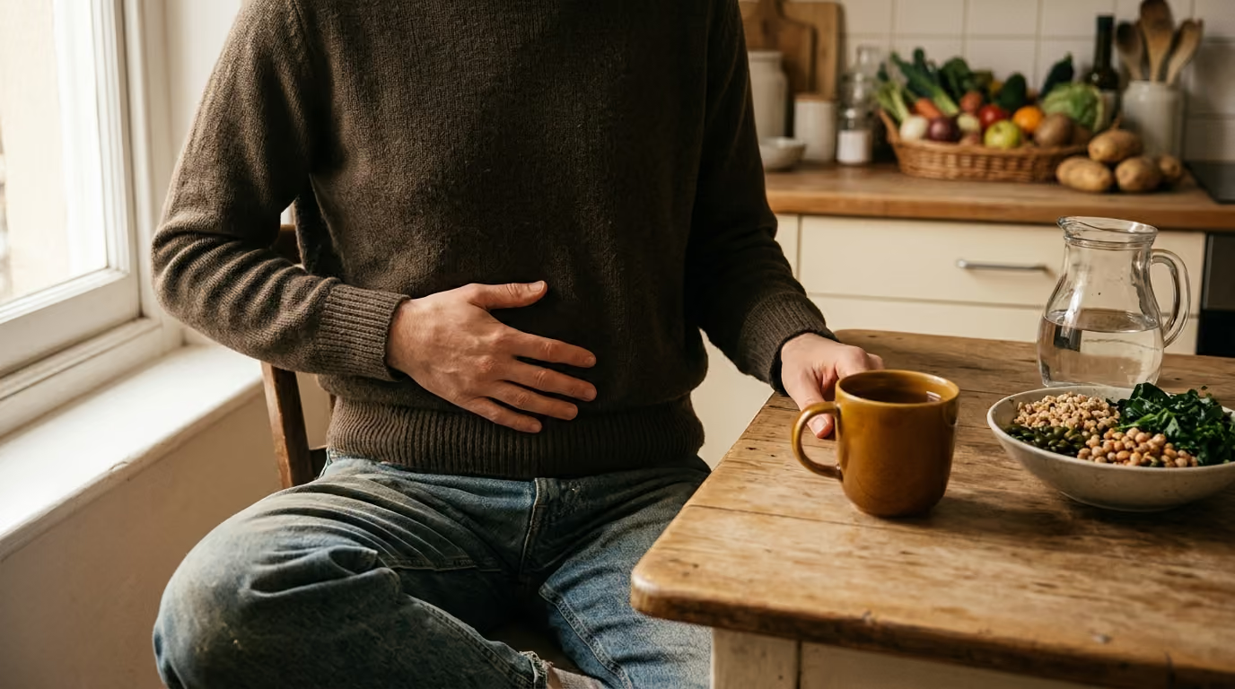

Three accessory digestive organs work together through coordinated secretions to break down fats, neutralize acids, filter bloodstream toxins, and protect overall metabolic health.





You sit down to enjoy a balanced dinner containing roasted chicken, vegetables, and olive oil. As you chew and swallow, your stomach begins churning the food into a liquid mixture called chyme.
Most people picture digestion as a simple conveyor belt running straight from the mouth to the colon. Yet the most intricate chemical transformations of your meal happen outside that main tube.
Tucked alongside your upper digestive tract sit three specialized structures: the liver, the gallbladder, and the pancreas. Food never actually passes through these organs. Instead, they act as an essential biochemical support network, manufacturing fluid, concentrating vital compounds, and neutralizing acids at precisely timed intervals.
Medical consensus recognizes the liver, gallbladder, and exocrine pancreas as the accessory digestive system. Working in tight coordination, they deliver specialized fluids into the small intestine that break down fats, carbohydrates, and proteins into absorbable nutrients. At the same time, this system protects the intestinal lining from harsh stomach acids and filters newly absorbed substances before they enter general circulation.
Understanding how these organs collaborate helps clarify how your body handles daily meals. It also sheds light on common digestive changes, from post-meal fullness to the effects of gallbladder surgery.
To understand digestion, it helps to distinguish the primary digestive tract from its accessory organs. The alimentary canal is a continuous muscular tube that extends from the mouth to the anus. In contrast, the liver, gallbladder, and pancreas are accessory organs located outside this pathway.
These organs connect to the digestive tract through a branching system of microscopic channels and larger ducts. Their shared biological mission can be understood through four core tasks: make, store, deliver, and process.
First, specialized cells in the liver and pancreas manufacture digestive secretions continuously or in response to biological signals. Second, the gallbladder stores and concentrates one of these vital fluids between meals. Third, a coordinated network of ducts delivers these fluids into the duodenum, which is the first segment of the small intestine. Fourth, the liver processes the nutrient-rich blood that returns from the intestines after digestion occurs.
The duodenum serves as the central meeting point for this entire accessory system. When acidic food leaves the stomach, it enters the duodenum through the pyloric sphincter. This triggers the release of bile from the biliary tract and pancreatic juice from the pancreas.
The anatomical route of these secretions requires precise alignment:
Because these pathways share a common drainage point, anatomical changes or physical blockages in one duct can influence the other. A blockage near this shared junction can disrupt both bile flow and pancreatic drainage at the same time. Learning about everyday gut function and motility demonstrates how tightly regulated these physical pathways truly are.
The liver is the largest internal organ in the human body, weighing roughly three pounds in an adult. While it performs hundreds of distinct metabolic tasks, its primary direct contribution to digestion is the continuous creation of bile.
Specialized liver cells called hepatocytes synthesize bile around the clock. Estimates of daily bile production vary across physiological studies based on measurement methods. Some clinical sources, such as the Merck Manual, estimate standard production at roughly 500 to 600 milliliters per day. Other references, including StatPearls, cite a range of 600 to 1,000 milliliters, while OpenStax notes it can reach roughly one full liter daily. Across all scientific literature, healthy adults produce roughly half a liter to one liter of bile every single day.
Bile is a complex, water-based fluid composed of several critical biological ingredients:
Beyond manufacturing bile, the liver acts as a critical processing facility for the entire digestive tract. This function relies on a unique vascular arrangement known as the dual blood supply.
Most organs receive blood solely from an artery and return it through a vein. The liver, however, receives oxygen-rich blood from the hepatic artery and nutrient-rich blood from the hepatic portal vein. The portal vein collects all the venous blood flowing away from the stomach, small intestine, large intestine, and spleen.
As a result, virtually every nutrient, medication, and compound absorbed through your intestinal lining must pass through the liver before reaching the rest of your body. Inside the liver, blood flows through microscopic channels called sinusoids. Hepatocytes sit immediately adjacent to these sinusoids, allowing them to survey, extract, and modify substances in the bloodstream.
When you consume a meal rich in carbohydrates, glucose enters the portal blood and travels straight to the liver. Hepatocytes remove surplus glucose from circulation and convert it into glycogen for short-term energy storage. When blood sugar drops between meals, the liver breaks down this stored glycogen and releases steady streams of glucose back into the blood.
The liver also alters potentially harmful byproducts of metabolism, neutralizes circulating chemicals, and prepares waste substances for excretion through bile or urine. This dual identity makes the liver both an upstream supplier of digestive secretions and a downstream processor of absorbed nutrients.
To understand why bile is necessary, consider the basic physical behavior of oil and water. The interior of the small intestine is a watery environment, while dietary fats are hydrophobic lipids that naturally clump together. Without an intermediary substance, dietary fats would remain in large, separate globules that digestive enzymes could not efficiently reach.
Bile salts solve this challenge through a physical process known as emulsification. Bile salts have an amphipathic chemical structure, meaning one end of the molecule is attracted to water while the other end is attracted to lipids.
When bile mixes with fats in the duodenum, the lipid-loving portions of the bile salts bury themselves inside the fat globules. The water-loving portions remain exposed on the outer surface facing the surrounding intestinal fluid.
This molecular arrangement breaks large fat masses into microscopic droplets, preventing them from coalescing back into large clumps. Emulsification does not chemically digest fat molecules on its own. Instead, it dramatically expands the surface area of the fat droplets, giving water-soluble pancreatic enzymes easy access to break the chemical bonds inside.
In addition to aiding fat breakdown, bile salts form tiny transport structures called micelles that assist in the absorption of fatty acids, monoglycerides, and fat-soluble vitamins (vitamins A, D, E, and K) across the intestinal wall.
Producing brand-new bile salts from cholesterol is an energy-intensive process for the liver. To conserve resources, your body uses an efficient recycling pathway known as enterohepatic circulation.
After bile salts complete their work in the upper small intestine, they travel downstream with the remaining digestive contents. When they arrive at the terminal ileum, which is the final section of the small intestine, specialized transport proteins actively reabsorb them into the bloodstream. According to medical references such as the Merck Manual, roughly 90 percent of bile salts are reabsorbed in this lower region.
These reclaimed bile salts enter the portal vein and travel directly back to the liver. Hepatocytes extract them from the blood and re-secrete them into fresh bile for use in future meals. The entire bile salt pool in your body recycles several times each day.
Other components of bile do not get recycled through this loop. Bilirubin, the yellow breakdown product from red blood cells, continues traveling past the ileum into the large intestine.
Once inside the colon, resident bacteria metabolize bilirubin into a series of secondary pigments, primarily stercobilin. Stercobilin is the natural compound responsible for giving human stool its characteristic brown color. When bile flow is blocked and cannot reach the gut, stool often turns a distinctive pale or clay-like shade due to the lack of this transformed pigment.
Readers interested in the broader biological impact of these secretions can read about gut microbiome and digestive science to see how bacterial activity continually modifies biliary compounds.
While the liver manufactures bile continually throughout the day and night, you only need large amounts of bile when you actually eat. The gallbladder serves as the storage reservoir that resolves this timing mismatch.
The gallbladder is a small, pear-shaped muscular sac nestled beneath the right lobe of the liver. It can hold roughly 30 to 60 milliliters of fluid in its resting state. Because liver bile is produced in much larger volumes, the gallbladder must do more than simply hold fluid; it actively concentrates it.
Between meals, the muscular sphincter at the entrance to the duodenum remains firmly closed. As the liver continues to secrete bile, the fluid backs up through the common bile duct and diverts through the cystic duct into the gallbladder.
Once inside, the inner epithelial lining of the gallbladder actively absorbs water and inorganic electrolytes like sodium and chloride from the fluid. According to OpenStax Anatomy and Physiology, this continuous absorption can concentrate bile salts, cholesterol, and pigments by up to tenfold.
During fasting periods, bile in the gallbladder becomes a dense, highly efficient fluid ready for rapid deployment. However, not all bile is stored exclusively in this reservoir. As noted in medical physiology texts, small amounts of dilute bile can also trickle directly from the liver into the small intestine between meals.
The signal to release concentrated bile begins when a meal leaves the stomach. When partially digested fats and proteins enter the duodenum, specialized endocrine cells in the intestinal wall release a hormone called cholecystokinin, commonly abbreviated as CCK.
Cholecystokinin travels through the bloodstream and exerts two coordinated physical effects:
This coordinated squeeze-and-release action squirts a concentrated bolus of bile through the cystic and common bile ducts directly into the duodenum, precisely when the arriving meal requires emulsification.
When the gallbladder is surgically removed due to gallstones or inflammation, a procedure known as a cholecystectomy, the body adapts to the structural change. The liver does not stop making bile, and bile ducts remain entirely functional.
Without the storage sac, bile can no longer be held and concentrated in advance of large meals. Instead, bile flows continuously from the liver through the bile ducts directly into the duodenum in a more dilute, steady stream. Over time, the common bile duct often dilates slightly to accommodate this steady flow, and most individuals continue digesting food normally.
The pancreas is an elongated, spongy gland that lies horizontally behind the stomach. It is a dual-purpose organ possessing two completely distinct physiological roles: an endocrine role and an exocrine role.
The endocrine pancreas consists of isolated cell clusters called the islets of Langerhans. These cells secrete hormones such as insulin and glucagon directly into the bloodstream to regulate systemic blood sugar levels.
In contrast, the exocrine pancreas makes up more than 95 percent of the organ's total tissue mass. Its sole responsibility is producing and delivering digestive fluids directly into the duodenal lumen. OpenStax estimates that the exocrine pancreas secretes over a liter of pancreatic juice every day.
Pancreatic juice is an alkaline fluid packed with two indispensable elements: digestive enzymes and bicarbonate ions. The production of these components is divided between two distinct cell types:
Bicarbonate plays a vital protective role in the upper digestive tract. Chyme leaving the stomach is extremely acidic, often possessing a pH between 1.5 and 2.5 due to gastric hydrochloric acid. If this acid entered the small intestine unchecked, it would rapidly erode the delicate intestinal lining and destroy digestive enzymes.
Pancreatic bicarbonate neutralizes this gastric acid on contact, raising the duodenal pH to a neutral or slightly alkaline range between 6.0 and 7.0. This neutralization halts the destructive activity of gastric pepsin and establishes the exact chemical environment required for intestinal and pancreatic enzymes to operate efficiently.
The exocrine pancreas manufactures enzymes specialized for every primary food group:
The handling of protein-digesting enzymes presents a biological dilemma: how does the pancreas manufacture potent proteases without digesting its own protein-rich cellular structure?
The pancreas solves this problem by synthesizing its proteases in inactive precursor forms known as zymogens or proenzymes. Acinar cells package these inactive zymogens into membrane-bound granules and secrete them into the ducts without active proteolytic activity.
The activation cascade begins only after the zymogens safely reach the interior of the duodenum:
An enzyme embedded directly in the duodenal brush-border membrane, called enteropeptidase, cleaves a small peptide segment from inactive trypsinogen. This converts it into active trypsin.
Once formed inside the intestinal cavity, active trypsin acts as the master switch, rapidly cleaving and activating the other resting zymogens, including chymotrypsinogen and procarboxypeptidase.
This multi-step safety mechanism keeps powerful protein digestion confined entirely to the intestinal lumen. If zymogens become prematurely activated inside the pancreas itself, the resulting self-digestion can lead to acute pancreatitis.
Just like the gallbladder, pancreatic secretion is closely regulated by meal-related hormones. When acidic chyme hits the duodenum, specialized endocrine cells release the hormone secretin. Secretin acts on pancreatic duct cells, stimulating them to flood the juice with water and bicarbonate.
Simultaneously, the presence of fats and protein fragments triggers the release of CCK. While CCK causes the gallbladder to contract, it also acts directly on pancreatic acinar cells, stimulating them to release their stored enzyme granules. Through the coordinated action of secretin and CCK, the small intestine receives exactly the combination of buffering fluid and digestive enzymes needed to process each specific meal.
Those seeking a deeper look at dietary balance can explore resources on nutrition, fiber, and gut-friendly eating to see how different macronutrients influence this digestive workflow.
The accessory digestive organs are frequently misunderstood in popular wellness discussions. Because their anatomy is complex and hidden from plain view, several persistent myths have developed around how they function.
One of the most widespread misunderstandings is the belief that the gallbladder produces bile. In reality, the gallbladder is entirely a storage, concentration, and delivery vessel.
Every drop of bile is manufactured upstream by hepatocytes inside the liver. The gallbladder simply receives this fluid between meals, removes excess water, and releases it when dietary fats stimulate contraction. When a person has their gallbladder removed, their liver continues to produce normal volumes of bile.
People often assume that bile breaks down fat molecules in the same chemical manner that stomach acid or enzymes break down food. However, bile salts contain no digestive enzymes whatsoever.
Bile acts strictly as a mechanical and physical emulsifier, breaking large lipid aggregates into microscopic suspensions. The actual chemical digestion of fats, cleaving triglyceride molecules into absorbable free fatty acids, is performed downstream by pancreatic lipase. Bile prepares the physical surface area so that lipase can do its chemical work.
It is common to imagine the liver as a large holding tank that collects and holds all the food you swallow. In reality, food never enters the liver.
The liver only interacts with food after it has been broken down into microscopic molecules and absorbed across the intestinal wall into the portal bloodstream. The liver then extracts, filters, and metabolizes these blood-borne nutrients, converting excess glucose to glycogen or building vital blood proteins from absorbed amino acids.
Because pancreatic juice is so effective at breaking down meals, some believe that all of its components are active enzymes. Bicarbonate is not an enzyme; it is an inorganic chemical buffer.
Bicarbonate does not break chemical bonds in nutrients. Instead, it neutralizes harsh stomach acids, raising the intestinal pH to create the neutral environment that allows other enzymes to operate.
Another misconception is that the body disposes of all bile salts after every meal. While certain components of bile like bilirubin are indeed waste products meant for elimination, bile salts are carefully preserved.
Through enterohepatic circulation, the body reclaims roughly 90 percent of its bile salt pool in the lower small intestine, returning those molecules to the liver to be used repeatedly throughout the week.
Popular wellness culture often promotes fasts, juices, or specialty tonics claiming to cleanse the liver, gallbladder, or pancreas. From a physiological standpoint, these organs do not accumulate layers of sludge that can be washed away by a beverage.
The liver continuously filters compounds using enzymatic pathways, while the gallbladder and pancreas regularly flush their ducts through normal hormonal contractions triggered by daily meals. Supporting these organs relies on balanced everyday nutrition and hydration rather than restrictive protocols.
Supporting the coordinated function of your liver, gallbladder, and pancreas does not require complex routines or restrictive diets. Because these organs respond directly to the composition and timing of your meals, simple and consistent dietary habits can promote smooth day-to-day operation.
One grounded, actionable step you can implement is ensuring a moderate, consistent distribution of dietary fats across your main meals rather than consuming very large fat loads in a single sitting.
When you eat meals that contain moderate amounts of healthy fats, such as olive oil, nuts, seeds, or avocado, the duodenum releases steady, measured pulses of CCK. This stimulates the gallbladder to contract completely and regularly, preventing concentrated bile from sitting stagnant for extended periods.
At the same time, moderate fat intake keeps the exocrine pancreas from being overwhelmed by a sudden, massive demand for lipase and bicarbonate. In contrast, skipping meals entirely followed by an unusually heavy, high-fat meal can place sudden mechanical and secretory demands on both the biliary ducts and the pancreatic acinar system.
Here are a few practical ways to establish this balanced meal rhythm:
For readers seeking to understand how lifestyle patterns influence broader digestive processes, our overview of the gut-brain connection and whole-body wellness offers additional context on how nervous system signals help regulate these secretions.
While the classic mechanical and enzymatic roles of the accessory organs have been understood for decades, contemporary research is uncovering a sophisticated bidirectional dialogue between these organs and the gut microbiome.
Historically, bile acids were viewed merely as biological detergents that assisted in fat digestion. Today, researchers recognize bile acids as potent signaling molecules that act throughout the entire body.
When bile salts enter the intestine, resident gut bacteria chemically alter them, converting primary bile acids made by the liver into secondary bile acids. These microbially modified bile acids bind to specialized receptors, such as the farnesoid X receptor (FXR) and the Takeda G-protein receptor 5 (TGR5).
These receptors sit on cells throughout the gut, liver, and immune system. When activated by modified bile acids, they send biochemical signals that help regulate liver lipid metabolism, maintain glucose homeostasis, and influence systemic inflammation.
Scientists are also investigating the bidirectional
communication between the exocrine pancreas and intestinal bacteria. Preliminary studies suggest that pancreatic juice contains natural antimicrobial peptides alongside its digestive enzymes. These peptides help shape the composition and spatial distribution of the bacterial community in the upper small intestine.
Conversely, microbial metabolites produced in the gut appear to communicate back to the pancreas through the bloodstream, potentially influencing how acinar and duct cells respond to dietary stimuli. While this research is still evolving, it highlights that the liver, gallbladder, and pancreas do not operate in biological isolation. Instead, they form an interactive network with the trillions of microorganisms residing within the gut lumen.
Because the liver, gallbladder, and pancreas share common physical pathways and perform vital metabolic tasks, structural blockages or tissue inflammation can produce distinct warning signs.
According to the National Institute of Diabetes and Digestive and Kidney Diseases (NIDDK), gallstones affect an estimated 10 to 15 percent of the United States population, representing nearly 25 million individuals. While many gallstones produce no symptoms, NIDDK notes that about a quarter of the nearly one million people diagnosed annually will require treatment, usually surgical intervention.
A primary risk associated with gallstones is their ability to migrate out of the gallbladder and lodge in shared ducts. If a gallstone blocks the common bile duct or the hepatopancreatic ampulla, it can obstruct both bile flow and pancreatic drainage. NIDDK identifies gallstone pancreatitis, an inflammation of the pancreas resulting from a gallstone blockage, as a serious condition that requires prompt hospital care.
You should consult a healthcare provider promptly if you notice any of the following symptoms:
These symptoms can point to acute inflammation of the gallbladder (cholecystitis), pancreatic inflammation (pancreatitis), or a biliary obstruction. A qualified medical professional can evaluate your symptoms using diagnostic tools such as blood chemistry panels, abdominal ultrasound, or specialized scans to accurately identify the underlying issue.
To learn more about everyday abdominal symptoms, our educational guide to bloating and regularity explains how intestinal motility patterns interact with normal digestion.
The liver, gallbladder, and exocrine pancreas demonstrate the remarkable coordination of the human body. Although food never travels through their physical tissues, their coordinated secretions make the digestion of every meal possible.
The liver continuously manufactures bile and filters every nutrient absorbed into the bloodstream. The gallbladder concentrates and stores this bile, releasing it on demand when fats enter the duodenum.
Simultaneously, the exocrine pancreas delivers a steady supply of neutralizing bicarbonate and inactive enzyme zymogens that dismantle starches, proteins, and lipids safely inside the intestinal lumen. Together, these organs form a balanced accessory system that ensures your body extracts essential nutrients while protecting the delicate lining of your gut.
When to revisit this resource: Refer back to this guide whenever you want to clarify how your digestive system handles fats and proteins, understand why gallbladder removal changes bile flow, or make sense of medical discussions regarding digestive enzymes and biliary health.
By looking past simplistic wellness claims and understanding the true anatomy of these accessory organs, you can make informed, grounded choices that support your daily digestive wellness.
DigestGenius publishes research-led guidance on digestion, the gut microbiome, fiber, probiotics, gut-brain signaling, inflammation and everyday digestive wellbeing.


Explore clear, research-led guidance on digestion, the microbiome, food, fiber, probiotics and the gut-brain connection.
read the blog