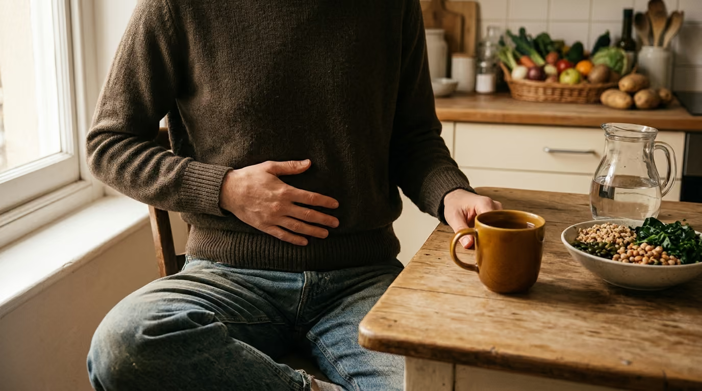

Post-meal digestive reflexes coordinate esophageal transit, stomach accommodation, fullness signaling, and bowel motility through automated neural and hormonal pathways.





You sit down to enjoy a relaxed dinner after a busy day. Within twenty minutes of finishing your plate, you feel a gentle fullness in your upper belly, release a quiet burp, and suddenly feel a distinct urge to visit the restroom. For many people, these rapid bodily shifts can feel surprising or even concerning. You might wonder if the meal moved through your body too quickly, or if your stomach is struggling to handle what you ate.
In reality, your digestive tract relies on a network of automated signals that activate the moment you begin eating. These coordinated responses, known as digestive reflexes, help your body receive food, manage internal pressure, and clear room along the gastrointestinal tract. Understanding how these biological programs function can help you separate everyday physiological events from patterns that deserve professional medical attention.
Medical researchers and gastroenterologists view post-meal digestion as an orchestrated physiological dialogue. The digestive tract does not operate as a passive chute where gravity does all the work. Instead, it functions as an active, sensory-rich muscular tube governed by the enteric nervous system, the central nervous system, and circulating gut hormones.
When food enters your mouth and travels toward your stomach, specialized mechanical and chemical sensors register its volume, temperature, and nutrient makeup. The scientific consensus is clear that the signals you notice after eating, such as stomach relaxation, gas venting, and shifts in lower bowel activity, are normal protective responses. The body uses these reflexes to accommodate incoming volume without causing sudden spikes in tissue tension.
These automated reflexes also prevent backflow of stomach contents, vent swallowed air, and prepare the lower intestines for upcoming digestive workloads. Because individual nerve sensitivity and gut motility vary naturally, two people can eat the exact same meal and experience entirely different internal sensations. Understanding these pathways through digestion and everyday gut function resources provides clarity on
why these signals occur.
The journey of any meal begins with swallowing, medically referred to as deglutition. While deciding to chew and initiate a swallow is voluntary, the subsequent transit down the esophagus is completely involuntary. Once a bolus of food passes the back of your throat, a coordinated neuromuscular wave takes over to guide it safely into the stomach.
The esophagus is a muscular corridor that connects the pharynx to the stomach. Its primary job is to transport swallowed solids and liquids while also clearing any material that might accidentally splash upward from below. When you swallow, the upper esophageal sphincter relaxes briefly to admit the food bolus.
Immediately afterward, a progressive contraction wave known as primary peristalsis sweeps down the length of the esophagus. This wave moves behind the bolus, gently propelling it downward. If any food particles linger along the esophageal lining, local stretch sensors trigger secondary peristalsis to sweep the corridor clean.
At the junction where the esophagus meets the stomach sits the lower esophageal sphincter, often abbreviated as the LES. Rather than acting like a rigid mechanical valve, the lower esophageal sphincter is a specialized ring of smooth muscle. It works in partnership with the crural diaphragm, which is the muscular portion of the diaphragm encircling the lower esophagus.
Together, the lower esophageal sphincter and the crural diaphragm create an active high-pressure barrier. When you swallow, the body must open this barrier to allow food to enter the stomach. Research on sphincter mechanisms shows that swallow-induced relaxation of the lower esophageal sphincter begins within about two seconds of swallowing and typically lasts between 6 and 10 seconds. Once the food bolus enters the stomach cavity, the sphincter muscle tightens again to restore the resting pressure barrier.
The lower esophageal sphincter does not only relax during a conscious swallow. It can also undergo what physiologists call transient lower esophageal sphincter relaxations, or TLESRs. These events occur independently of swallowing and are completely normal occurrences in healthy individuals.
During a transient relaxation, the sphincter relaxes for a longer duration, often lasting several seconds more than a typical swallow-induced relaxation. This opening allows the stomach to vent accumulated air and balance internal pressures. Recognizing that the sphincter is designed to open and close dynamically helps clarify that intermittent relaxation is an essential physiological function rather than an inherent structural failure.
Once food clears the lower esophageal sphincter, it enters the stomach, which serves as both a chemical processing vat and an adaptable storage reservoir. The stomach must handle varied meal sizes without letting internal pressures rise to uncomfortable or damaging levels.
To accommodate an incoming meal, the stomach uses two distinct yet complementary physiological mechanisms: receptive relaxation and gastric accommodation. Receptive relaxation occurs almost instantaneously as you swallow. Signals traveling along the vagus nerve tell the proximal stomach, which includes the fundus and upper body, to soften its muscular tone in anticipation of incoming food.
Gastric accommodation, on the other hand, is the sustained adaptation of the upper stomach as meal volume progressively increases. In healthy digestion, the muscular walls of the fundus and gastric body relax and stretch, increasing their compliance. This adaptive compliance allows the stomach to hold a substantial volume of food and beverage without a parallel surge in internal pressure. Under ordinary physiological conditions, this smooth expansion happens automatically without causing conscious distress.
This remarkable elasticity is regulated primarily by the vagovagal reflex, a communication loop involving the vagus nerve and the brainstem. Mechanoreceptors embedded within the stomach wall detect the stretching caused by food. These receptors send sensory signals up to the central nervous system, which then sends inhibitory motor signals back to the smooth muscle of the gastric fundus.
These inhibitory signals prompt the release of neurotransmitters, such as nitric oxide and vasoactive intestinal peptide, which command the stomach muscle fibers to lengthen and relax. When this pathway operates smoothly, you can finish a complete meal while experiencing a calm, comfortable sense of nourishment rather than abrupt mechanical pressure.
As eating continues and the stomach approaches its physical capacity, the nature of internal sensory signaling changes. While initial stretch prompts relaxation, continued distension eventually crosses a biological threshold. Once this threshold is reached, stretch receptors along the gastric wall send strong afferent signals to the brain, contributing directly to the sensation of fullness and satiety.
These same signals begin to stimulate rhythmic contractions in the lower stomach, known as the antrum. The antrum acts as a muscular mill, grinding solid food particles down into a fine semi-liquid mixture called chyme. Learning more about digestive health fundamentals helps contextualize how stomach relaxation and antral grinding work together during a meal.
One of the most common post-meal experiences is feeling an urge to have a bowel movement shortly after finishing food. For many people, this signal appears within ten to thirty minutes after breakfast or lunch. This sudden sensation is governed by a well-documented physiological mechanism known as the gastrocolic reflex.
The gastrocolic reflex is an automated physiological response where food entering and stretching the stomach stimulates increased motility in the large intestine. When the stomach wall expands to receive a meal, it sends rapid forward signals to the colon. The purpose of this reflex is straightforward: it encourages the colon to clear out older, processed waste material to make room for the new nutrients currently entering the upper digestive tract.
This reflex is entirely physiological, meaning its presence is a normal sign of an active, communicating nervous system. Experiencing this post-meal urge does not mean that your digestive system is dysfunctional or overly sensitive. It simply demonstrates that your upper and lower digestive organs are sharing information effectively.
Clinical research shows that colonic motility can increase within minutes of food entering the stomach. A person may notice a gentle urge to pass stool within minutes to roughly an hour after eating, and this heightened colonic activity can persist for up to a few hours.
The motor response in the colon involves both an increase in baseline muscular tone and periodic bursts of high-amplitude contractions. These powerful waves are known as mass movements. Mass movements sweep through the transverse and descending colon, shifting existing fecal matter into the sigmoid colon and rectum. Once stool reaches the rectum, local stretch receptors trigger the conscious sensation that it is time to use the restroom.
The gastrocolic reflex does not rely on a single biological switch. Instead, it involves an integrated network of autonomic nerves, the enteric nervous system, and several key biochemical messengers. When food contacts the stomach and upper small intestine, specialized endocrine cells release signaling molecules into the bloodstream and local tissues.
Key mediators involved in this response include:
Because dietary fat is a particularly potent trigger for cholecystokinin release, richer meals often prompt a noticeably stronger gastrocolic response than lighter meals. Readers navigating these daily patterns can find deeper context in our guide to bloating and regularity.
The act of eating involves not only the intake of food and fluids, but also the inevitable intake of ambient air. Managing this trapped air requires coordinated venting mechanisms involving the upper stomach, the lower esophageal sphincter, and the diaphragm.
Every time you swallow food, drink through a straw, or talk while eating, you swallow small quantities of air, a process known as aerophagia. The oxygen and nitrogen in swallowed air cannot be completely absorbed across the stomach lining. As a result, this gas gathers at the top of the stomach fundus, forming a gas bubble.
As this gas pocket grows, it gently distends the upper gastric wall. In response, mechanoreceptors trigger a transient lower esophageal sphincter relaxation. This temporary opening allows the gas bubble to escape upward into the esophagus, where it travels safely to the pharynx and is released as a burp. Gastric belching is therefore a protective venting mechanism that prevents excess gas pressure from building up in the stomach cavity.
The diaphragm is widely recognized as the primary muscle of respiration, but it also plays a vital mechanical role in digestive comfort. The esophagus passes through an opening in the diaphragm called the esophageal hiatus. The muscular fibers surrounding this opening, known as the crural diaphragm, wrap around the lower esophageal sphincter like an external collar.
During times of increased abdominal pressure, such as when you bend over, laugh, or take a deep breath, the crural diaphragm contracts firmly. This pinch-cock action reinforces the lower esophageal sphincter, preventing acidic stomach contents from splashing upward into the chest. The intimate partnership between breathing musculature and esophageal tissue highlights how deeply interconnected your structural anatomy is with digestive function.
Another diaphragm-related reflex that can appear around mealtime is the hiccup, known clinically as singultus. A hiccup consists of a sudden, involuntary contraction of the diaphragm and intercostal muscles, followed immediately by abrupt closure of the vocal cords, which produces the characteristic sound.
Post-meal hiccups are often mistakenly blamed on a simple stomach spasm, but the reflex arc is far more complex. Afferent signals that trigger hiccups can originate from sensory nerve endings in the distal esophagus, the stomach wall, or the abdominal side of the diaphragm itself. These sensory messages travel along the phrenic nerve, the vagus nerve, and sympathetic nerve chains to the brainstem. Eating too quickly, consuming carbonated beverages, or experiencing sudden temperature changes in the stomach can stimulate these sensitive nerve pathways, triggering a brief series of hiccups until the nerve loop calms down.
While digestive reflexes are typically silent and comfortable, the body also possesses powerful protective motor programs designed to defend against perceived threats. Nausea, retching, and vomiting are sophisticated, highly coordinated physiological responses rather than random muscular failures.
Nausea is an unpleasant subjective sensation that often signals a sudden change in upper gastrointestinal motor activity. When nausea develops, the normal rhythmic contractions of the stomach fundus and body diminish rapidly, leading to a loss of baseline gastric tone. At the same time, the resting pressure of the pyloric sphincter, which guards the exit from the stomach into the small intestine, decreases.
These motor shifts cause the stomach to act as a relaxed, passive receptacle. This sudden cessation of downward motility prepares the upper digestive tract for a potential reversal of flow if the central nervous system determines that ingested material must be expelled.
Retching represents a rhythmic, coordinated muscular event that frequently precedes vomiting. During retching, the respiratory muscles of the chest wall, the abdominal wall muscles, and the diaphragm contract simultaneously against a closed airway.
These opposing muscular contractions create massive pressure fluctuations between the chest cavity and the abdominal cavity. The diaphragm contracts forcefully downward while the abdominal muscles squeeze inward, raising intra-abdominal pressure and pushing stomach contents upward into the lower esophagus. However, because the upper esophageal sphincter remains tightly closed during retching, the gastric contents slide back down into the stomach between contractions.
Vomiting is the final expulsive phase where gastric and upper intestinal contents are forcefully projected out through the mouth. Unlike a simple muscular contraction of the stomach wall, vomiting is driven by external skeletal muscles.
During the expulsive phase, the abdominal wall muscles contract intensely, and the diaphragm undergoes a rapid, coordinated shift in motor activity to allow pressure transmission. The lower esophageal sphincter, the esophagus, and the upper esophageal sphincter all relax completely. The elevated pressure inside the abdominal cavity then propels the stomach contents smoothly upward through the wide-open esophageal corridor and out of the body. Understanding these integrated steps shows that expulsion is a meticulously managed protective reflex coordinated by the brainstem.
Because post-meal sensations happen inside the body where they cannot be seen, it is easy to misinterpret what normal signals mean. Clarifying these widespread misunderstandings can ease unnecessary health worries and promote a calmer relationship with your body.
Many people who feel an immediate urge to use the restroom after eating believe their digestive tract works like a straight, open pipe. They assume that the food they just swallowed raced through their stomach and intestines in less than half an hour.
In reality, standard gastrointestinal transit takes anywhere from 24 to 72 hours under healthy conditions. The urge you feel twenty minutes after eating is the gastrocolic reflex at work. The stretching of your stomach simply sends an electrical and hormonal message to your colon, encouraging it to evacuate waste from meals eaten one or two days prior.
Another frequent assumption is that releasing a burp after eating is an early indicator of gastroesophageal reflux disease, commonly known as GERD. While individuals with GERD can certainly experience frequent belching, the act of burping by itself does not establish a medical diagnosis.
Gastric belching is the primary biological method your body uses to vent swallowed air. Releasing air through transient sphincter relaxations prevents excess pressure from building inside the stomach cavity. Unless belching is accompanied by persistent burning pain, regurgitation of acidic liquid, or difficulty swallowing, it is typically an ordinary venting reflex.
It is tempting to picture healthy digestive sphincters as rigid, permanently sealed valves that only open when food pushes past them. Following this logic, any relaxation that occurs without food passing downward is viewed as a defect.
Human anatomy relies on dynamic, flexible muscular barriers. The lower esophageal sphincter is programmed to relax during conscious swallowing and during transient venting episodes. These relaxations allow gas to escape and prevent painful gastric over-distension. A healthy sphincter is defined by its ability to coordinate opening and closing appropriately, not by remaining clamped shut at all times.
When people notice a strong sense of fullness after a moderate meal, they sometimes worry that their stomach has stopped working or that food is sitting motionless in their gut. This worry can create anxiety around future meals.
Feeling full is a normal sensory milestone generated by gastric accommodation and subsequent stretch-receptor activation. As the stomach fundus expands to receive a meal, it eventually sends signals along the vagus nerve to let your brain know you have consumed enough energy. A sensation of comfortable fullness indicates that your stretch pathways and vagal communication loops are functioning as designed.
Many popular explanations treat post-meal hiccups as an isolated muscular twitch occurring entirely within the stomach wall. This oversimplification overlooks the complex neural circuitry involved in the response.
A hiccup is a coordinated reflex involving the diaphragm, the intercostal muscles of the ribs, the vocal cords, and three major nerve networks. Sensory input from the esophagus, stomach, or diaphragmatic peritoneum travels to the central nervous system, which coordinates the sudden motor response. Recognizing this multi-structure reflex provides a far more accurate picture of how your body manages sudden shifts in thoracic and abdominal volume.
While your body manages digestive reflexes automatically, everyday habits can significantly influence how smoothly these reflexes operate. Applying grounded, sensible lifestyle habits can reduce unnecessary gastrointestinal strain and help you feel more comfortable after eating.
One of the most effective ways to support digestive harmony is to adjust the pace of your meals. Eating too rapidly causes you to swallow substantial amounts of excess air, which quickly distends the stomach fundus. This rapid stretching can trigger frequent transient sphincter relaxations, leading to excessive belching, hiccups, or uncomfortable early fullness.
Take time to chew your food into a fine, manageable consistency before swallowing. Chewing thoroughly lubricates the food bolus with saliva, which contains initial digestive enzymes like salivary amylase. Smaller, well-lubricated bites pass easily through the esophagus, allowing the lower esophageal sphincter to relax and close in a smooth, unhurried rhythm.
Your physical posture during and immediately after eating has a direct mechanical effect on gastric accommodation and reflux prevention. Slouching or hunching over a desk compresses the abdominal cavity, artificially raising intra-abdominal pressure. This external pressure can push stomach contents against the lower esophageal sphincter, challenging the natural barrier created by the crural diaphragm.
Instead, sit upright while eating and remain in an upright or gently reclined position for at least thirty to sixty minutes afterward. Keeping your torso elongated gives your stomach ample room to expand outward during gastric accommodation. Taking a gentle, unhurried ten-minute walk after eating can also support natural stomach emptying and encourage balanced motility without putting stress on your digestive tract.
How you structure your meals can also influence the intensity of your post-meal reflexes. Consuming extremely large, high-fat meals triggers a massive release of cholecystokinin and gastrin, which can cause an overly intense gastrocolic reflex or delayed stomach emptying. Balancing your plate with modest portions of healthy fats, lean proteins, and soluble fiber supports steady gastric processing.
Additionally, pay attention to how you consume fluids around mealtime. Gulping large glasses of ice-cold water or carbonated drinks while eating can rapidly distend the stomach and trigger sudden temperature changes that stimulate hiccup pathways. Sipping room-temperature beverages in moderate quantities keeps your digestive tissues comfortable and allows gastric juices to interact effectively with your food. Exploring our resources on gut-brain lifestyle connections offers further ideas for creating relaxing mealtime routines.
Gastroenterology research is continuously expanding our understanding of how the nervous system and the gut communicate. While the core mechanics of swallowing, accommodation, and the gastrocolic reflex are well established, current studies are exploring why digestive reflexes feel so different from one person to another.
One active area of investigation centers on visceral hypersensitivity, a condition where the sensory nerve endings in the digestive tract become unusually reactive to normal physiological events. In people with heightened visceral sensitivity, routine stomach stretching during gastric accommodation can be perceived as intense pressure or pain, even when internal pressures remain completely normal.
Researchers are studying how low-grade mucosal inflammation, alterations in the gut microbiome, and central nervous system processing contribute to this heightened sensory state. Understanding these pathways helps explain why some individuals experience postprandial distress syndrome, a subtype of functional dyspepsia characterized by bothersome post-meal fullness or early satiety, despite having normal stomach anatomy.
Physiologists are also refining the tools used to measure how the stomach adapts to food. Historically, assessing gastric compliance required an invasive procedure known as a gastric barostat, which uses an inflatable balloon placed in the stomach to measure volume and pressure changes.
Modern clinical researchers are validating non-invasive alternatives, including specialized nutrient drink challenge tests, high-resolution ultrasound, and advanced MRI imaging. These emerging diagnostic modalities allow doctors to visualize how the fundus expands and how the crural diaphragm moves during real-time digestion. As these tools become more widely accessible, they will provide deeper insights into individual variations in gastric tone and post-meal motor reflexes.
Everyday digestive reflexes are a normal part of life, but persistent or severe symptoms should never be ignored. While an occasional burp, a brief bout of hiccups, or a post-breakfast urge to use the restroom are ordinary occurrences, recurring discomfort warrants an individualized assessment by a qualified healthcare professional.
Gastroenterologists look for specific clinical warning signs, often referred to as alarm features, to determine when post-meal symptoms require structured diagnostic testing, such as an upper endoscopy or imaging studies.
You should consult a physician if you experience any of the following concerning symptoms:
If you find yourself frequently distressed by post-meal fullness, severe bloating, chronic diarrhea, or painful heartburn, avoid trying to self-diagnose based on reflex descriptions alone. Many gastrointestinal conditions share overlapping symptoms, and an accurate diagnosis requires a thorough review of your medical history, dietary habits, and overall health.
A primary care physician or a gastroenterologist can perform targeted evaluations, rule out underlying organic diseases, and develop a personalized care plan tailored to your specific physiology. Working alongside a medical professional ensures you receive safe, evidence-based guidance while supporting your long-term digestive wellbeing. To learn more about our educational philosophy, visit our about page.
You may want to revisit this guide whenever you experience shifts in your mealtime routine, notice changes in how your body responds to different foods, or find yourself feeling anxious about ordinary bodily noises and sensations. Returning to these core anatomical explanations can help ground your perspective in solid digestive physiology.
By viewing your body as a coordinated biological system rather than a collection of confusing symptoms, you can interpret post-meal signals with calm confidence and make informed choices for your everyday health.
DigestGenius publishes research-led guidance on digestion, the gut microbiome, fiber, probiotics, gut-brain signaling, inflammation and everyday digestive wellbeing.


Explore clear, research-led guidance on digestion, the microbiome, food, fiber, probiotics and the gut-brain connection.
read the blog